Class 11 - Biology
Chapter 6 - Anatomy of Flowering Plants
Top Block 1
Question 1.State the location and function of different types of meristems.
Answer :
Meristems are of three types on the basis of their location in plant body:
(i) Apical meristem: It is present at the apices of root and shoot and is responsible for increase in length.
(ii)Intercalary meristem: It is present at the bases of leaves above the nodes or below the nodes and is responsible for elongation of the organs.
(iii)Lateral meristem : It is present on lateral side and is responsible for increase in girth or diameter.
Question 2.Cork cambium forms tissues that form the cork. Do you agree with this statement? Explain.
Answer :
Yes, I agree with this statement. Cork cambium cuts off cells both on its outer side and inner side. The cells cut off on outer side form cork and cells cut off on inner side form secondary cortex. The cells of cork are dead whereas those of secondary cortex are living.
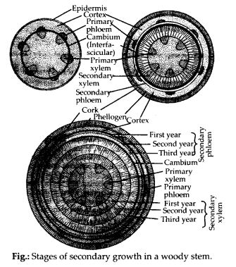
Question 3.Explain the process of secondary growth in the stems of woody angiosperms with the help of schematic diagrams. What is its significance?
Answer :
Secondary growth is the formation of secondary tissues from lateral meristems. It is found in dicots only. It increases the diameter of the stem. Secondary tissues are formed by two types of lateral meristems, vascular cambium and cork cambium. Vascular cambium produces secondary vascular tissues while cork cambium forms periderm.The vascular bundles in dicot stem are conjoint, collateral, open and are arranged in a ring. The cambium present between xylem and phloem in vascular bundles is called fascicular or intrafascicular cambium. Besides this, some cells of medullary rays also become meristematic and this is called interfascicular cambium. Both these cambia collectively constitute complete ring of vascular cambium. This ring of vascular cambium divides periclinally to cut off cells both on inner side and outer side. The cells cuts off on outer side are secondary phloem and inner side are secondary xylem. Amount of secondary xylem cut off is more than secondary phloem and thus with the formation of secondary tissue, increase in girth or diameter occurs. The structure of secondary xylem and secondary phloem is similar to that of primary xylem and primary phloem. With the increase in secondary tissue, the primary xylem and primary phloem get crushed. The ray initials of vascular cambium ring divide by tangential divisions and add new cells. These new cells produced on both the sides of ray initials remain meristematic for sometime and then differentiate into parenchymatous cells of rays. The rays, produced by vascular cambium in between the secondary xylem and secondary phloem, are called secondary medullary rays. They are usually one to few layers in thickness and one to several layers in height. The medullary rays form the radial systejn responsible for radial conduction of solutes. They maintain connection between pith and cortex There is a marked difference in activity of cambium with change in season. In spring, the activity of cambium is more and hence the wood elements are larger in size with wide lumen. The activity of cambium is less during autumn and the wood elements are smaller in size with narrow lumen. Spring wood and autumn wood of a year constitute annual ring.
In order to increase in girth and prevent harm on the rupturing of the outer ground tissues due to the formation of secondary vascular tissues, dicot stems produce a cork cambium or phellogen in the outer cortical cells. Phellogen cells divide on both the outer side as well as the inner side to form secondary tissues. The secondary tissue formed on the inner side is called secondary cortex while the tissue formed on outer side is called cork.
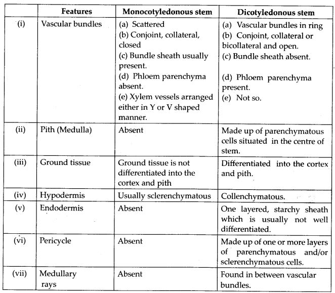
Question 4.Draw illustrations to bring out the anatomical difference between
(a) Monocot root and dicot root
(b) Monocot stem and dicot stem
Answer :
(a) Differences between monocot root and dicot root are illustrated in the following figure and table.


Mddle block 1
(b) Differences between monocot and dicot stems are illustrated in the following figure and table.

Question 5.Cut a transverse section of young stem of a plant from your school garden and observe it under the microscope. How would you ascertain whether it is a monocot stem or a dicot stem ? Give reasons.
Answer :
Vascular bundles in dicot stem are arranged in a ring whereas in monocot stem vascular bundles are scattered throughout the ground tissue. On the basis of arrangement of vascular bundles it can be ascertained
whether the young stem is dicot or monocot. Besides undifferentiated ground tissue, sclerenchymatous hypodermis, oval or circular vascular bundles with Y shaped xylem are other differentiating features of monocot stem.
Question 6.The transverse section of a plant material shows the following anatomical features – (a) the vascular bundles are conjoint, scattered and surrounded by a sclerenchymatous bundle sheath, (b) phloem parenchyma is absent. What will you identify it as?
Answer :
The plant material is identified as monocot stem.
Question 7.Why are xylem and phloem called complex tissues?
Answer :
A group of different types of cells which perform common function is called complex tissue. Xylem and phloem are called complex tissues as all cells that work as a unit for a common function have different structural organisation. Xylem has four types of cells-tracheids, vessels, xylem parenchyma and xylem fibres. Phloem consists of sieve tube elements, companion cells, phloem parenchyma and phloem fibres. Xylem is associated with conduction of water and minerals from roots to top of plants and phloem is responsible for transport of organic food.
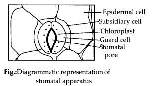
Question 8.What is stomatal apparatus? Explain the structure of stomata with a labelled diagram.
Answer :
Stomata are structures present in the epidermis of leaves. Stomata regulate the process of transpiration and gaseous exchange. Each stoma is composed*of two bean shaped cells known as guard cells which enclose stomatal pore. The outer walls of guard cells (away from the stomatal pore) are thin and the inner walls (towards the stomatal pore) are highly thickened. The guard cells possess chloroplasts and regulate the opening and closing of stomata. Sometimes, a few epidermal cells, in the vicinity of the guard cells become specialised in their shape and size and are known as subsidiary cells. The stomatal aperture, guard cells and the surrounding subsidiary cells are together called stomatal apparatus.
Question 9.Name the three basic tissue systems in the flowering plants. Give the tissue names under each system.
Answer :
The three basic tissue systems in flowering plants are epidermal tissue system, ground tissue system and vascular tissue system.
Epidermal tissue system comprises epidermal cells, stomata, trichomes and hairs.
Ground tissue system consists of cortex, endodermis, pericycle, pith and medullary rays, in the primary roots and stems. In¬leaves, the ground tissue consists of thin walled chloroplast containing cells and is called mesophyll.
The vascular tissue system consists of complex tissues, the phloem and the xylem.
Question 10.How is the study of plant anatomy useful to us?
Answer :
Study of internal structures of plants is called plant anatomy. Study of plant anatomy is useful:
-for solving taxonomic problems.
-for knowing homology and analogy of various plant groups.
-to differentiate the superior and inferior, standard and substandard or specified and unspecified woods.
-in establishing purity and correct identity of plant parts in pharmacognosy (science connected with sources, characteristics and possible medicinal uses).
-in knowing the structural peculiarities of different groups of plants.
Question 11 .What is periderm? How does periderm formation take place in the dicot stems?
Answer :
phelloderm, phellogen and phellem together constitute the periderm. Periderm is protective in function.Dicot stems produce cork cambium or phellogen in the outer cortical cells. Phellogen cells divide on both the outer side as well as the inner side to form secondary tissues. The secondary tissue produced on the inner side of the phellogen is called secondary cortex or phelloderm. On the outer side phellogen produces cork or phellem.
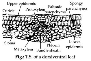
Question 12.Describe the internal structure of a dorsiventral leaf with the help of labelled diagram.
Answer :
Bottom Block 3
Click here to visit Official CBSE website
Click here for NCERT solutions
Click here to visit Official Website of NCERT
Click here to download NCERT Textbooks