Class 11 - Biology
Chapter 7 - Structural Organisation in Animals
Top Block 1
Question 1. Answer in one word or one line.
(i) Give the common name of Periplaneta americana.
(ii) How many spermathecae are found in earthworm?
(iii) What is the position of ovaries in cockroach?
(iv) How many segments are present in the abdomen of cockroach?
(v) Where do you find Malpighian tubules?
Answer :
(i) Cockroach.
(ii) Four pairs.
(iii) In cockroach two large ovaries, lie laterally in the 2nd – 6th abdominal segments’.
(iv) Abdomen of cockroach consists of 10 segments.
(v) Malpighian tubules are present at the junction of midgut and hindgut in cockroach.
Question 2. What are the following and where do you find them in animal body?
(a) Chondrocytes
(b) Axons.
(c) Ciliated epithelium
Answer :
(a) Chondrocytes – Chondrocytes are the only cells found in cartilage. They are present in spaces called lacunae and they produce and maintain the matrix of cartilage. Bending ability of cartilage is due to chondrocytes. Cartilage is present at tip of nose, pinna of ear, epiglottis etc.
(b) Axon – Axon is one of the processes of neuron, which is the structural and functional unit of nervous system. The part of cyton – n’here axon arises is axon hillock and axon ends in group of branches called terminal arborizations. It conducts impulses away from the cyton. Neurons (nerve cells)
are present in brain and spinal cord.
(c) Ciliated epithelium – If the columnar or cuboidal cells bear cilia on their free surface they are called ciliated epithelium. Their function is to move particles or mucus in a specific direction over the epithelium. They are mainly present in the inner surface of hollow organs like bronchioles and Fallopian tube.
Question 3. Draw a labelled diagram of the reproductive organs of an earthworm.
Answer :

Question 4. Answer the following.
(i) What is the function of nephridia?
(ii) How many types of nephridia are found in earthworm based on their location?
Answer :
(i) Nephridia are excretory organs of earthworm, which perform the function of excretion and osmoregulation. Nephridia regulate the volume and composition of the body fluids. A nephridium is a coiled tubular and microscopic structure which starts out as a funnel that collects excess fluid from coelomic chamber. The funnel connects with a tubular wastes through a pore to the surface in the body wall or into the digestive tube.
(ii) In earthworm, nephridia are present in all segments except the first two. There are three types of nephridia on the basis of their location:
(a) Septal nephridia, present on both the sides of intersegmental septa from segment 15 to the last that open into intestine.
(b) Integumentary nephridia, attached to lining of the body wall of segment 3 to the last that open on the body surface and
(c) Pharyngeal nephridia, present as three paired tufts in the 4th, 5th and 6th segments.

Mddle block 1
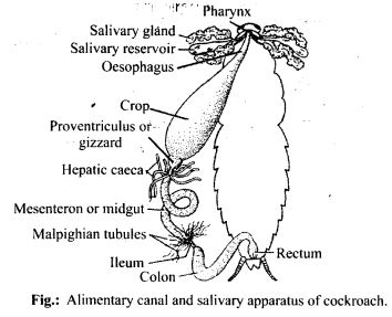
Question 5. Draw a labelled diagram of alimentary canal of a cockroach.
Answer :
Question 6. What are the cellular components of blood?
Answer :
Blood is a fluid connective tissue. It is composed of plasma (fluid) and blood cells (corpuscles). Cellular components of blood (blood corpuscles) constitute about 45% of blood volume.
Three types of blood cells are:
(i) Erythrocytes or red blood cells: They are most abundant blood cells. Normal RBC count is 5-5.5 million/mm3 in males and 4.5-5 million/mm3 in females) RBCs help in transport of gases and maintain blood pH.
(ii) Leucocytes or white blood cells: The normal WBC count is 5000-6000/mm3 of blood. They are involved in immune response of body and act as soldiers and scavangers.
(iii) Thrombocytes or blood platelets: There are about 2,50,000 platelets/mm3 of blood. They are involved in blood clotting.
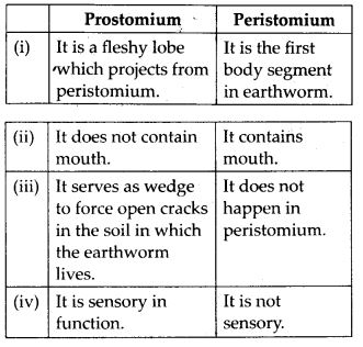
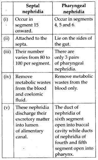
Question 7. Distinguish between the following:
(a) Prostomium and peristomium
(b) Septal nephridium and pharyngeal
Answer :
(a) Differences between prostomium and peristomium are
Question 8. Mark the odd one in each series.
(a) Areolar tissue; blood; neuron; tendon
(b) RBC; WBC; platelets; cartilage
(c) Exocrine; endocrine; salivary gland; ligament
(d) Maxilla; mandible; labrum; antennae
(e) Protonema; mesothorax; metathorax; coxa.
Answer :
(a) Neuron: Areolar tissue, blood and tendon are connective tissues while neuron is a part a nervous tissue.
(b) Cartilage: RBC, WBC and platelets are parts of vascular connective tissue while cartilage is skeletal connective tissue.
(c) Ligament: Ligament is a connective tissue.
(d) Antennae: Maxilla, mandible and labrum are mouth parts of cockroach while antennae are sense organs.
(e) Protonema: Protonema is a filamentous juvenile stage in life cycle of Bryophytes, while mesothorax, metathorax and coxa are appendages of cockroach.
Question 9. Match the terms in column I with those in column II.
Column I | Column II |
| (a) Compound epithelium (b) Compound eye (c) Septal nephridia (d) Open circulatory system (e) Typhlosole (f) Osteocytes (g) Genitalia | (i) Alimentry canal (ii) Cockroach (iii) Skin (iv) Mosaic vision (v) Earthworm (vi) Phallomere (vii) Bone |
Solution: (a) – (iii), (b) – (iv), (c) – (v), (d) – (ii), (e) – (i), (f) – (vii), (g) – (vi)
Question 10. Mention briefly about the circulatory system of earthworm.
Answer :
Earthworm possesses a closed type of blood vascular system, as the blood flows through closed blood vessels. Blood is red in colour due to respiratory pigment haemoglobin. Prominent blood vessels in earthworm includes dorsal, ventral, sub- neural, lateral oesophageal and supra- oesophageal blood vessels. There are four pairs of tubular hearts, provided with valves. The anterior two pairs of hearts, known as lateral hearts lie in the 7th and 9th segments and connect the dorsal blood vessel with the ventral blood vessel. They receive blood from the dorsal blood vessel and convey it to the ventral blood vessel. The posterior two pairs of hearts are called latero-oesophageal hearts and are situated in the 12th and 13th segments. The latero-oesophageal hearts apart from connecting the dorsal and ventral blood vessels are also joined with the supra oesophageal blood vessel. Latero-oesophageal hearts carry blood from the dorsal vessel and the supra oesophageal vessel to the ventral blood vessel.Contractions keep blood circulating in one direction. Blood glands are present in the 4th, 5th and 6lh segments which produce blood cells and haemoglobin which is dissolved in blood plasma. Blood cells are phagocytic in nature.
Question 11. Describe various types of epithelial tissues with the help of labelled diagrams.
Answer :
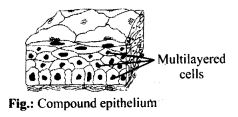
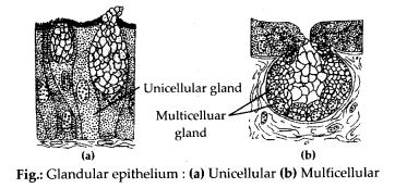
Epithelial tissue is a tissue made of one or more layers of compactly arranged cells that covers external surface and internal free surface of body organs and which is underlined by a basement membrane. The various types of epithelial tissue along with the diagram are given below:
(i) Simple epithelium : It is composed of single layer of cells which rest on basement membrane. Simple epithelium generally occurs over secretory and absorptive surfaces and forms lining of body cavities, ducts and tubes. Simple epithelium is of several types.
(a) Squamous epithelium: It consists of single layer of flat cells, tightly linked together and have centrally located oval or spherical nucleus. It is also called pavement epithelium. It is found in walls of blood vessels, air sacs of lungs, and lining of eye lens.
(b) Cuboidal epithelium: Cells of cuboidal epithelium are as tall as wide, with centrally placed nucleus. Its main functions are secretion and absorption. It lines sweat gland, thyroid follicles, salivary glands. Brush bordered cuboidal epithelium, i.e., cells having microvilli on their free surface lines proximal part of uriniferous tubule, pancreatic duct, testis and ovary.
(c) Columnar epithelium: Cells are with basally located nucleus. It helps in secretion and absorption. It occurs in lining of intestine, stomach, gall bladder.
(d) Ciliated epithelium: Free surface of columnar and cuboidal cells are covered with cilia. Cilia help in moving fluids, particles, mucus, etc. in a specific direction. It occurs in the inner surface of Fallopian tubules, nasal passage, bronchioles.


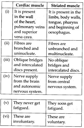
Question 12. Distinguish between
(a) Simple epithelium and compound epithelium.
(b) Cardiac muscle and striated muscle.
(c) Dense regular and dense irregular connective tissues.
(d) Adipose and blood tissue.
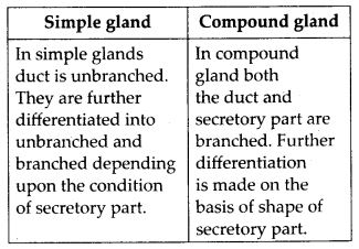
(e) Simple gland and compound gland.
Answer :
(a) Differences between simple and compound epithelium are as follows:


Question 13. Draw a neat diagram of digestive system of frog.
Answer :

Question 14. Mention the function of the following:
(a) Ureters in frog
(b) Malpighian tubules
(c) Body wall in earthworm.
Answer :
(a) Ureters in frog: Ureter is a transparent duct which arise from outer portion of kidney. In the “male frogs, ureter acts as urinogenital duct which runs backwards from kidneys and opens into the cloaca. It carries both urine and spermatozoa from kidney to the cloaca. In female, ureter conducts only urine from kidneys to the cloaca.
(b) Malpighian tubules: Malpighian tubules are excretory organs present in cockroach. These are present at junction of mid gut and hindgut. These are fine, long, unbranched, yellowish and blind tubules and are 100-150 in number. They help in the removal of excretory products from haemolymph.
(c) Body wall in earthworm: It consists of cuticle, epidermis, muscular layer and parietal peritoneum.
(i) It maintains the characteristic shape of’ the body.
(ii) It protects the internal organs.
(iii) The cuticle prevents excessive evaporation.
(iv) It serves as an ideal respiratory organ.
(v) The receptor cells play a vital sensory function.
(vi) The albumen helps in the formation of cocoon. It also serves as a food for the developing earthworm inside the cocoon.
(vii) Setae and muscles are responsible for locomotion.
(viii) Excretory matter is passed out through nephridiopores.
Bottom Block 3
Click here to visit Official CBSE website
Click here for NCERT solutions
Click here to visit Official Website of NCERT
Click here to download NCERT Textbooks